

Most Important Biology Diagram For NEET UG 2026: Biology diagrams are among the most powerful scoring tools in the NEET UG exam, and mastering them can quickly push your score toward the 650+ range.
In Day 10 of our “Most Important Biology Diagram” series, we focus on a high-yield figure that is repeatedly tested through direct questions, labelling, and concept-based MCQs.
Understanding not just the structure but also the function and clinical relevance behind each labelled part will help you revise faster, remember longer, and attempt questions with complete confidence.

Sagittal Section of Human Brain
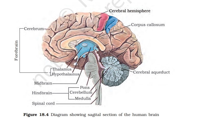
1. Cerebrum
- Largest part of the brain (forebrain).
- Responsible for thinking, intelligence, memory, learning, emotions, and voluntary actions.
- Controls sensory perception like vision, hearing, smell, and touch.
2. Cerebral Hemisphere
- Two halves of the cerebrum (left and right hemispheres).
- Connected by the corpus callosum.
- Show functional specialization (e.g., left → language, right → creativity/spatial skills).
3. Corpus Callosum
- Thick band of nerve fibers connecting the two hemispheres.
- Allows communication and coordination between left and right brain.
4. Thalamus
- Acts as a sensory relay station.
- Receives sensory impulses and forwards them to the cerebrum.
- Important for pain, touch, temperature, and awareness.
5. Hypothalamus
- Maintains homeostasis.
- Controls:
- Body temperature
- Hunger & thirst
- Sleep and emotions
- Endocrine system via pituitary gland
6. Midbrain
- Connects forebrain and hindbrain.
- Controls visual and auditory reflexes.
- Helps regulate eye movement and posture.
7. Pons
- Bridge between different brain parts.
- Helps regulate breathing rhythm.
- Transfers signals between cerebrum and cerebellum.
8. Cerebellum
- Responsible for balance, posture, and coordination of muscles.
- Ensures smooth and precise voluntary movements.
- Important for motor learning (e.g., cycling, writing).
9. Medulla Oblongata
- Controls vital involuntary functions:
- Breathing
- Heartbeat
- Blood pressure
- Swallowing, coughing, vomiting
- Damage here can be life-threatening.
10. Cerebral Aqueduct
- Narrow canal connecting third and fourth ventricles.
- Allows circulation of cerebrospinal fluid (CSF) in the brain.
11. Spinal Cord
- Continuation of the medulla.
- Conducts nerve impulses between brain and body.
- Responsible for reflex actions.
Read Also: Most Important Biology Diagram For NEET UG 2026 (Day 9)
PRACTICE QUESTIONS
Q.1. In a strict mid-sagittal section, which ventricular structure is most clearly visible along the midline?
A. Fourth ventricle
B. Lateral ventricle (frontal horn)
C. Third ventricle
D. Cerebral aqueduct
Q.2. Which structure is best described as a large C-shaped bundle of white matter clearly seen in a mid-sagittal section?
A. Cingulate gyrus
B. Corpus callosum
C. Superior sagittal sinus
D. Internal capsule
Q.3. In a mid-sagittal section, where is the primary location of the pineal gland relative to surrounding structures?
A. Anterior to the hypothalamus and above the optic chiasm
B. Embedded in the cingulate gyrus
C. Inferior to the medulla and dorsal to the spinal cord
D. Between the superior colliculi and the splenium of the corpus callosum
Q.4. On a mid-sagittal section, which structure forms the inferior and anterior walls of the third ventricle?
A. Hypothalamus
B. Thalamus
C. Hippocampus
D. Epithalamus
Q.5. In a mid-sagittal section, which structure directly connects the third ventricle to the fourth ventricle?
A. Central canal
B. Cerebral aqueduct
C. Interventricular foramen
D. Foramen magnum
Human Fetus Within Uterus
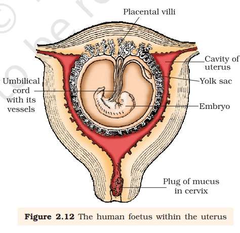
1. Placental villi
Feature:
- Finger-like projections of the placenta that extend into maternal uterine tissue.
- Rich in foetal blood capillaries and surrounded by maternal blood spaces.
Function:
- Site of exchange of oxygen, nutrients, CO₂, and wastes between mother and foetus.
- Also helps in hormone secretion (hCG, hPL, estrogens, progesterone, relaxin).
2. Umbilical cord with its vessels
Feature:
- Connects the foetus to the placenta.
- Contains two umbilical arteries and one umbilical vein embedded in Wharton’s jelly.
Function:
- Umbilical vein: carries oxygenated, nutrient-rich blood to the foetus.
- Umbilical arteries: carry deoxygenated blood and wastes to the placenta.
3. Cavity of uterus
Feature:
- Hollow muscular chamber lined by endometrium.
- Contains amniotic fluid and developing embryo/foetus.
Function:
- Provides space, protection, and nourishment for foetal development.
- Uterine muscles help in labour and childbirth.
4. Yolk sac
Feature:
- Small membranous sac attached to the embryo in early development.
- Prominent in early weeks of pregnancy.
Function:
- Site of early blood cell formation.
- Supplies initial nutrients before placenta becomes fully functional.
5. Embryo (developing foetus)
Feature:
- Early developmental stage after implantation.
- Gradually differentiates into organs and body systems.
Function:
- Undergoes growth, organogenesis, and maturation to become a foetus and later a newborn.
6. Plug of mucus in cervix
Feature:
- Thick mucus seal formed in the cervical canal during pregnancy.
Function:
- Prevents entry of microbes into the uterus.
- Maintains a sterile and protected environment for the foetus.
- Expelled near time of delivery.
PRACTICE QUESTIONS
Q.1. In a typical placenta diagram, chorionic villi extend into pools of maternal blood. What is the main functional role of these villi?
A. To pump maternal blood back toward the uterine arteries
B. To store nutrients and oxygen for later release to the fetus
C. To mix fetal blood directly with maternal blood for faster exchange
D. To greatly increase the surface area for diffusion between maternal blood and fetal capillaries
Q.2. In a labeled placenta diagram, maternal blood is shown filling the intervillous space, while fetal capillaries run inside the villi. Which statement best describes the relationship between these two blood supplies?
A. Maternal blood remains confined inside maternal capillaries located within the villi
B. Fetal blood flows through the uterine arteries and veins along with maternal blood
C. Maternal blood remains outside the villi and bathes them, while fetal blood stays inside capillaries within the villi
D. Maternal and fetal blood mix freely in the intervillous space to form a shared circulation
Q.3. On a placenta diagram, the umbilical cord connects the fetus to the placenta. Which best describes the role of the umbilical cord vessels in relation to the placental villi and maternal blood?
A. The umbilical cord vessels transport fetal blood to and from the placenta, where it passes through capillaries in the villi near maternal blood
B. The umbilical cord vessels pump oxygen directly into the fetal lungs, bypassing the placenta
C. The umbilical cord vessels carry maternal blood from the uterus into the fetal heart
D. The umbilical cord vessels carry both maternal and fetal blood together through a single shared vessel
Q.4. In a schematic of the uterine wall and placenta, maternal blood is shown entering the intervillous space from uterine arteries. What is the functional significance of this arrangement around the villi?
A. It prevents maternal blood from coming near the villi, limiting nutrient exchange
B. It ensures maternal blood flows in a closed loop inside fetal capillaries
C. It directs maternal blood only to the umbilical cord, bypassing the placental surface
D. It creates a low-resistance pool where maternal blood can slow down and bathe many villi simultaneously.
Consistent diagram practice is the secret weapon of NEET toppers, and adding just one important figure to your revision every day can create a huge difference over time. Make sure you revise today’s diagram, test yourself on the labels, and connect it with NCERT concepts for maximum retention.
Read Also: NEET UG Registration 2026: Documents Required & Eligibility Explained
Stay tuned for Day 11 in this series, where we’ll break down another must-know Biology diagram to keep your preparation sharp, focused, and exam-ready until NEET UG 2026.
Answers & Explanations
Ans.1. D. To greatly increase the surface area for diffusion between maternal blood and fetal capillaries. In the human placenta, chorionic villi are finger-like projections that arise from the fetal chorion. They extend into the intervillous spaces (pools/lacunae filled with maternal blood). Each villus contains fetal blood capillaries inside it.The primary functional role of these villi is to dramatically increase the surface area available for exchange of materials (oxygen, nutrients, carbon dioxide, and waste products) between the mother’s blood and the fetus’s blood.
Ans.2. C. Maternal blood remains outside the villi and bathes them, while fetal blood stays inside capillaries within the villi. Maternal blood is delivered into the intervillous spaces (also called lacunae) through the spiral arteries of the uterus.These intervillous spaces are large, blood-filled pools outside the chorionic villi.The chorionic villi (finger-like projections from the fetal side) project into these pools, so they are bathed by maternal blood.Inside each chorionic villus, there are fetal capillaries carrying fetal blood.Thus, maternal blood remains outside the villi (in the intervillous space), while fetal blood remains inside the capillaries within the villi.
Ans. 3. A. The umbilical cord vessels transport fetal blood to and from the placenta, where it passes through capillaries in the villi near maternal blood
Ans. 4. D. It creates a low-resistance pool where maternal blood can slow down and bathe many villi simultaneously.
ALL THE BEST!
Answers please
updated in post, kindly refer to it.
Daily practice 2026 UG neet mock test kindly helf me team
Go to the NEET UG Resources section on the edufever website. There you will find all the quizzes. (https://www.edufever.com/tag/neet-ug-quizzes/)