
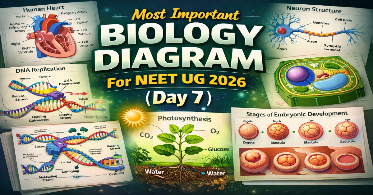
Most Important Biology Diagram For NEET UG 2026: Preparing for NEET UG 2026 is not just about reading chapters- it’s about mastering the concepts that frequently appear in the exam. Biology diagrams play a crucial role in helping you visualize processes, remember structures faster, and score easy marks with confidence. In Day 7 of our Most Important Biology Diagram series, we bring you another high-yield diagram that strengthens your conceptual clarity and boosts your chances of reaching the 650+ score target.
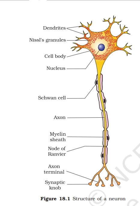
NEURON
A neuron is the basic functional unit of the nervous system, specialized for receiving, processing, and transmitting information via electrical and chemical signals.
Key Points To Remember
- Cell body (soma): Central part containing the nucleus and organelles; integrates signals.
- Dendrites: Branched extensions that receive incoming signals from other neurons.
- Axon: Long, slender projection that transmits signals away from the cell body.
- Myelin sheath: Fatty insulating layer around the axon that speeds up signal conduction.
- Nodes of Ranvier: Gaps in the myelin sheath enabling saltatory conduction for faster impulses.
- Axon terminals: End branches releasing neurotransmitters at synapses.
- Nissl’s granules: are basophilic clusters of rough endoplasmic reticulum (RER) and free ribosomes found in the cytoplasm of the neuronal cell body (soma) and dendrites.
- Nucleus: The nucleus resides in the neuron’s cell body (soma), housing genetic material (DNA) that directs protein synthesis, cell growth, and maintenance, often near Nissl granules for ribosomal support.
- Schwann cells: are glial cells in the peripheral nervous system that form the myelin sheath around axons, insulating them to speed electrical impulses via saltatory conduction between nodes of Ranvier.
- Synaptic knobs: are bulbous axon terminal ends that store neurotransmitters in vesicles, releasing them across the synaptic cleft to transmit signals to the next neuron or target cell.
Read Also: NCERT Diagrams for NEET UG 2026: The Most Underrated NEET Scoring Tool

PRACTICE QUESTIONS
Q.1. In a diagram of a typical multipolar neuron, which labelled part should be identified as the main site of impulse conduction away from the cell body?
A. Dendrites
B. Nissl bodies
C. Node of Ranvier
D. Axon
Q.2. A labelled diagram shows a neuron with the cell body at one end, a single long process in the middle, and synaptic terminals at the opposite end. Which label should represent the region rich in Nissl bodies and the nucleus?
A. Cell body (soma)
B. Node of Ranvier
C. Dendritic tip
D. Axon terminal
Q.3. In a diagram comparing a myelinated and an unmyelinated axon, which labelled feature is directly responsible for saltatory conduction in the myelinated neuron?
A. Myelin sheath with nodes of Ranvier
B. Schwann cell nucleus
C. Continuous plasma membrane of the axon
D. Axon hillock
Q.4. A labelled diagram shows a neuron whose cell body is located in the dorsal root ganglion, with a single process that later branches into two. Which type of neuron does this most likely represent?
A. Multipolar neuron
B. Bipolar neuron
C. Pseudo-unipolar neuron
D. Non-myelinated interneuron
Q.5. In a diagram of a synapse between two neurons, which labelled region should be identified as the synaptic cleft?
A. The narrow gap between pre- and postsynaptic membranes
B. The cytoplasm of the presynaptic terminal
C. The postsynaptic receptor region on the dendrite
D. The vesicle-filled region of the axon terminal
Read Also: NEET UG 2026: Re-Reading is not Revision, How NEET Revision Actually Works
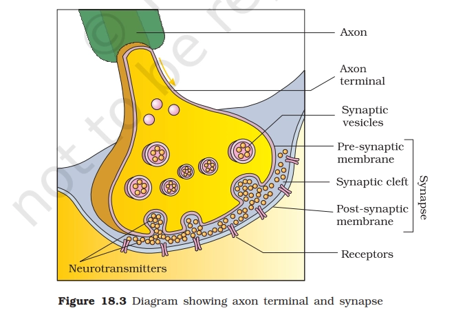
Axon Terminals and Synapses
Key Points To Remember
- Synaptic cleft: Narrow gap (20-40 nm) between presynaptic axon terminal and postsynaptic membrane.
- Postsynaptic receptors: On the target cell, bind neurotransmitters to propagate or inhibit signals.
- Pre-synaptic Membrane: This is the specialized plasma membrane at the axon terminal’s end, featuring the active zone where synaptic vesicles dock and fuse to release neurotransmitters upon calcium influx from arriving action potentials.
- Receptors: Primarily located on the post-synaptic membrane, these protein structures bind neurotransmitters diffused across the synaptic cleft, triggering ion channel opening or signaling cascades to propagate (excitatory) or inhibit (inhibitory) the signal.
- Axon Terminal: The bulbous distal axon end (synaptic knob) housing vesicles, mitochondria, and voltage-gated calcium channels.
PRACTICE QUESTIONS
Q.6. At a chemical synapse between two neurons, which event occurs immediately after an action potential reaches the axon terminal of the presynaptic neuron?
A. Reuptake of neurotransmitter into the presynaptic terminal
B. Opening of ligand-gated ion channels on the postsynaptic membrane
C. Generation of an action potential in the postsynaptic neuron
D. Fusion of synaptic vesicles with the presynaptic membrane
Q.7. Which structure is directly responsible for storing neurotransmitters in the axon terminal of a neuron?
A. Mitochondria
B. Schwann cells
C. Synaptic vesicles
D. Nissl bodies
Q.8. In a typical cholinergic synapse, what primarily determines whether the postsynaptic membrane will generate an action potential?
A. The diameter of the synaptic vesicles in the axon terminal
B. The speed of axonal conduction in the presynaptic neuron
C. The number of mitochondria present in the axon terminal
D. The sum of excitatory and inhibitory postsynaptic potentials reaching threshold
Answers & Explanations
Ans.1. D. Axon. The axon is the long, cylindrical process that carries nerve impulses away from the cell body toward another neuron, muscle, or gland. It usually arises from the axon hillock and ends in terminal boutons or synaptic knobs.
Ans.2. A. Cell body (soma). The soma encloses the nucleus and most of the rough endoplasmic reticulum (Nissl bodies), which carry out the synthesis of proteins essential for neuronal repair, membrane maintenance, and neurotransmitter production.
Ans.3. A. Myelin sheath with nodes of Ranvier. Saltatory conduction occurs because myelin-covered segments of the axon prevent ion exchange, and voltage-gated channels cluster at the nodes.
Ans.4. C. Pseudo-unipolar neuron. These neurons have a single process that splits into two branches, giving a T-shaped appearance in diagrams.
Ans.5. A. The narrow gap between pre- and postsynaptic membranes. The synaptic cleft is a tiny extracellular space, typically about 20–40 nm wide, that separates the presynaptic membrane of the axon terminal from the postsynaptic membrane of the next cell.
Ans.6. D. Fusion of synaptic vesicles with the presynaptic membrane. Following the arrival of an action potential at the presynaptic terminal, voltage-gated channels open, allowing entry.
Ans.7. C. Synaptic vesicles. Located inside the axon terminal, synaptic vesicles store neurotransmitter molecules until an action potential triggers their fusion with the presynaptic membrane.
Ans.8. D. The sum of excitatory and inhibitory postsynaptic potentials reaching threshold. The postsynaptic cell integrates all the EPSPs and IPSPs it receives, and only if the net effect depolarizes the membrane to threshold will an action potential be initiated.
hii! can I get the answers, please?
The Answers and Explanations have been uploaded. Kindly refer to the post again.