
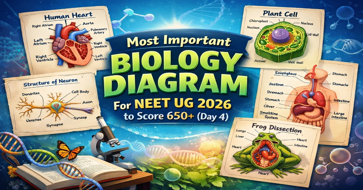
Most Important Biology Diagram For NEET UG 2026: What if scoring extra marks in NEET UG 2026 Biology was as simple as remembering a picture? NCERT diagrams are like hidden treasure- clear, direct, and often asked in the exam. Welcome to Day 4 of the Most Important Biology Diagrams series. Mastering this single diagram can turn an easy question into guaranteed marks, and we’re here to make it simple, visual, and actually fun to remember.
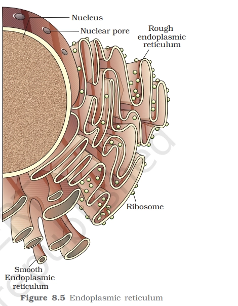
ENDOPLASMIC RETICULUM
The Endoplasmic Reticulum is a network of membrane-bound tubules and flattened sacs present in the cytoplasm of eukaryotic cells. It plays a central role in protein synthesis, lipid production, and intracellular transport.
Key Points to Remember:
While revising the ER diagram from NCERT, focus on these must-remember labels:

- Nuclear membrane connection – ER is continuous with the outer nuclear membrane.
- Ribosomes on Rough ER (RER) – Small dots attached to the surface.
- Smooth tubular network of Smooth ER (SER) – Lacks ribosomes.
- Lumen (internal cavity) – Space inside the ER where proteins/lipids are processed.
- Transport vesicles – Budding vesicles carrying materials to the Golgi apparatus.
- Rough Endoplasmic Reticulum (RER)
- Ribosomes attached to the surface.
- Synthesizes secretory and membrane proteins.
- Prominent in protein-secreting cells (e.g., plasma cells).
- Smooth Endoplasmic Reticulum (SER)
- No ribosomes present.
- Responsible for lipid synthesis, steroid formation, and detoxification.
- Well developed in liver cells and muscle cells.
Read Also: Most Important Biology Diagram For NEET UG 2026 (Day 3)
Practice Questions:
Q.1. In the given diagram, what is true for both A and B?
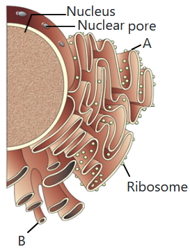
A. They are involved in protein synthesis.
B. They synthesize steroidal hormones in animal cells
C. They are a part of endomembranous system of a eukaryotic cell
D. They are continuous with the inner membrane of the nucleus
Q.2. In a typical diagram of a eukaryotic cell, which feature best helps you identify rough endoplasmic reticulum (RER) rather than smooth endoplasmic reticulum (SER)?
A. Tubular network without attached particles
B. Flattened cisternae studded with small dots
C. Large spherical body with double membrane
D. Stacked, curved sacs with distinct cis and trans faces
Q.3. In a labeled diagram of the endoplasmic reticulum, which region is most directly involved in the synthesis of proteins destined for secretion outside the cell?
A. Free ribosomes in the cytosol
B. Inner membrane of the mitochondrion
C. Lumen of the smooth ER
D. Ribosomes attached to the rough ER membrane
Read Also: Most Important Biology Diagram For NEET UG 2026 to Score 650+ (Day 2)
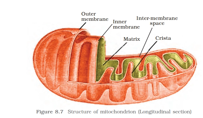
MITOCHONDRIA
Key Points To Remember
- Each mitochondrion is a double membrane-bound structure with the outer membrane and the inner membrane dividing its lumen distinctly into two aqueous compartments i.e., the outer compartment and the inner compartment.
- The inner compartment is filled with a dense homogeneous substance called the matrix.
- The inner membrane forms several infoldings called the cristae (sing.: crista)
- Mitochondria are the sites of aerobic respiration. They produce cellular energy in the form of ATP,
- The matrix also possesses
- single circular DNA molecule.
- Few RNA molecules,
- Ribosomes (70S) and the components required for the synthesis of proteins.
Practice Questions
Q.4. In a labeled mitochondrion diagram, which numbered structure would most likely represent the matrix?
A. The space between the outer and inner mitochondrial membranes
B. The highly folded inner membrane itself
C. A central, fluid-filled region enclosed by the inner membrane
D. The smooth outer membrane surrounding the organelle
Q.5. In a diagram showing a mitochondrion cut open, which label most likely points to the cristae?
A. The narrow region between the inner and outer membranes
B. The finger-like folds projecting inward from the inner membrane
C. The smooth, outermost boundary of the organelle
D. The small dots inside the matrix representing ribosomes
Q.6. Which labeled component of a mitochondrion would you associate with the presence of its own DNA and ribosomes?
A. Inner membrane
B. Matrix
C. Intermembrane space
D. Outer membrane
Q.7. A diagram shows arrows pointing to small granular structures within the mitochondrial matrix. What are these most likely representing?
A. Lysosomes
B. Peroxisomes
C. Golgi vesicles
D. Mitochondrial ribosomes
DNA REPLICATION FORK
For long DNA molecules, since the two strands of DNA cannot be separated in its entire length (due to very high energy requirement), the replication occur within a small opening of the DNA
helix, referred to as replication fork.
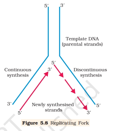
Key Points To Remember
- DNA-dependent DNA polymerases, many additional enzymes are required to complete the process of replication with high degree of accuracy.
- The DNA-dependent DNA polymerases catalyse polymerisation only in one direction, that is 5′ – 3′.
- On one strand (the template with polarity 3′-5′), the replication is continuous, while on the other (the template with polarity 5′-3′), it is discontinuous.
- The discontinuously synthesised fragments are later joined by the enzyme DNA ligase.
- There is a definite region in E. coli DNA where the replication originates. Such regions are termed as origin of replication.
Practice Questions
Q.8. Where would you locate DNA ligase on a detailed replication fork diagram, and what is its role?
A. Ahead of the fork, unwinding DNA and preventing supercoiling.
B. Coating single-stranded DNA to prevent re-annealing.
C. On the leading strand near the origin, starting DNA synthesis de novo.
D. Between adjacent Okazaki fragments on the lagging strand, forming phosphodiester bonds.
Q.9. In a labeled prokaryotic replication fork, which feature helps you identify the leading strand specifically?
A. It has multiple RNA primers spaced along its length.
B. It is synthesized in short fragments away from the replication fork.
C. It is associated only with DNA polymerase I and DNA ligase.
D. It shows a single continuous arrow of DNA synthesis in the same direction as fork movement.
Q.10. A replication fork with several labeled arrows along the lagging strand, each pointing away from the fork. What do these arrows most likely represent?
A. Flow of genetic information during transcription.
B. Direction of helicase movement separating the strands.
C. Movement of DNA ligase joining fragments towards the fork.
D. Direction of Okazaki fragment synthesis by DNA polymerase III.
One diagram, a few smart tricks, and you’re already closer to your 650+ NEET goal. That’s the power of learning Biology the visual way- quick to revise, easy to recall, and perfect for exam pressure moments. Keep coming back to this daily diagram journey, because every day adds one more sure-shot concept to your scorecard.
Comment Below for Answers and Explanations!!
All The Best!
Looking for answer
Q.1. C.
Q.2. B. flattened cisternae studded with small dots. In a diagram, rough endoplasmic reticulum is usually drawn as a series of flattened membrane sacs (cisternae) with numerous tiny dots on the outer surface. These dots represent ribosomes attached to the cytosolic side of the RER membrane.
Q.3. D. ribosomes attached to the rough ER membrane. Rough ER is characterized by ribosomes on its cytosolic face, and these ribosomes perform protein synthesis for secretory and membrane proteins.
Q.4. C.
Q.5. B
Q.6. B
Q.7. D
Q.8. D
Q.9. D.
Q.10.D.
Good questions but where are the answers?
Q.1. C.
Q.2. B. flattened cisternae studded with small dots. In a diagram, rough endoplasmic reticulum is usually drawn as a series of flattened membrane sacs (cisternae) with numerous tiny dots on the outer surface. These dots represent ribosomes attached to the cytosolic side of the RER membrane.
Q.3. D. ribosomes attached to the rough ER membrane. Rough ER is characterized by ribosomes on its cytosolic face, and these ribosomes perform protein synthesis for secretory and membrane proteins.
Q.4. C.
Q.5. B
Q.6. B
Q.7. D
Q.8. D
Q.9. D.
Q.10. D.